The Mowat Lab studies the senses (vision, hearing, olfaction) and the brain, in health, aging, and disease.
We study the effects of aging on sensory and cognitive health. Our work has direct relevance to dog and human age-related sensory loss including age-related retinal degeneration and age-related macular degeneration.
As a veterinary ophthalmologist and a researcher, Dr. Mowat is interested in the similarities between vision in dogs and humans and what predisposes the retina of both species to disease and degeneration. We have shown that there are substantial parallels between dog and human retinal anatomy, and visual aging. Our research also studies how visual decline impacts aging and decline of other senses and the brain. We use a translational approach to study how aging of companion dogs and humans that cohabit compares. Our studies are designed to create, optimize, and utilize sensory outcome measures for companion dogs that have direct parallels to human outcome measures.
***RECRUITING NOW!****
Does your dog have cataracts?
Are you planning and/or scheduling cataract surgery for your dog?
If yes, read on!
If you have cataract surgery performed for your dog, you can join an exciting new study that will allow us to learn more about the benefits for dogs of performing cataract surgery, particularly older/elderly dogs.
Is my dog eligible?
Your dog must have cataract surgery with a veterinary ophthalmologist planned in the near future (you must have completed a consultation with a veterinary ophthalmologist, and have surgery planned within 3 months). Surgery must be planned to be performed in at least one eye, and your dog must have significant vision impairment before surgery. You must have lived with your dog for at least 3 months, and you must plan to live with them for at least 6 months after surgery.
What is involved?
We will ask you to fill out standardized questionnaires about your dog, and the health of their vision, hearing, thinking/memory skills, behavior and quality of life (15-20 minutes to complete). We will invite you to complete one questionnaire before cataract surgery is completed, and 6 months after surgery.
Are there any benefits to me?
You and your dog will not receive any direct benefit. You will receive a $10 gift card (digital delivery) when you complete the study questionnaires.
Are there any risks?
We do not forsee any risks to your and your dog. There is a small risk that your identifiable information (name, home address, email, telephone number) becomes know to someone outside the study. We have rigorous security protocols in place to limit that risk.
How do I sign up?
If you would like to express interest in the study, please go to the AVMA clinical trials registry page, and complete the qualification survey (green button): https://veterinaryclinicaltrials.org/study/VCT24005825 A study team member will review your responses and contact you to invite you to participate, if you and your dog are eligible.
What if I have more questions?
Please reach out to Dr. Mowat (Study principal investigator) at mowat@wisc.edu if you have more questions.
VISION IN DOGS
The Canine “Macula”
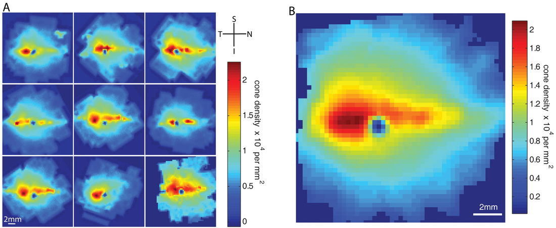
Dr. Mowat has worked to develop a deep understanding of the architecture of the central retina of dogs.
As humans, we have a specialized region of extremely high cone photoreceptor density, known as the fovea, where most of our “quality” visual tasks are focused, for example, reading and color perception. Dogs do not possess a traditional fovea but have a region of enriched cone photoreceptor density, known as the area centralis. This region may vary in location and prominence between different breeds of dogs.
Dog Screen Interaction Project
We recently completed a community science project asking the question “how do dogs interact with screens in the home”? The reason for this study is twofold:
a. To learn more about dog behavior. We leave our dogs alone in the home a lot, and is the “TV content” built for dogs actually something dogs like to engage in?
b. To learn more about dog vision. As dog vision researchers we want to be able to design an “eye chart” (similar to the one you use at the eye doctor yourself) for dogs, but in order to keep dogs engaged, we need to provide interesting content. What is that content and how long can we expect it to be engaging for? We have no idea, and this research will get us closer to understanding what dogs like to look at on screens!
The survey is now closed for participation and we thank those that participated!
Here is a link to the published study.
Highlights of our findings include:
• Age and visual status influence owner-perceived dog interactions with screens in the home
-
- younger dogs and dogs with better vision are more likely to interact with screens)
• Sporting and herding dog breeds watch more than other breed types.
• Movement on screens is a strong motivator for screen attention in dogs.
• Animal content is broadly interesting to dogs, with dog subjects the most popular.
• Dogs frequently exhibit signs of excitement when reacting to screen-based content.
Here is one of our lab members’ dogs watching TV in the lab!
We are still working in this space!
The Mowat lab is developing non-invasive methods to study vision and visually-mediated behavior in dogs, in addition to other sensory functions. We are doing this using a combination of dog owner questionnaires, and behavioral tests.
The Wisconsin Aging and Geriatric Dog (WAGD) study ![]()
We are currently recruiting dog participants for an exciting study of aging in current Beaver Dam Offspring Study-Neurocognitive Aging Study (BOSS-NCAS) and Wisconsin Sleep Cohort (WSC) participants’ dog families! Dog cohabiting BOSS-NCAS and WSC participants may receive an introduction/invitation letter in the coming months, containing a survey to complete about contact preferences so that the WAGD study team can contact you about recruiting your dog. Please reach out to dogaging@vetmed.wisc.edu with any questions! Unfortunately we are not able to recruit dogs outside of the BOSS-NCAS and WSC studies at this time.
Understanding why the macula area is susceptible to aging and disease.
The fovea and surrounding macula of people is photoreceptor dense and at risk of certain devastating blinding diseases such as age-related macular degeneration (AMD) and macular dystrophy. We study the unique susceptibility of the canine area centralis to disease, to better define why the human macula might be at risk of disease. We also study risk factors that combine with aging (diet, genetics) to increase the risk of retinal dysfunction. The ultimate goal is to find vision sparing solutions for human sufferers of blinding diseases such as AMD. We are particularly interested in the role of mitochondrial health in aging and disease of the retina, particularly the importance of a master regulator of mitochondrial function – peroxisome proliferative activated receptor gamma, coactivator 1-alpha (PPARGC1a or PGC1a).
SARDS
What is SARDS?
SARDS, also known as Sudden Acquired Retinal Degeneration Syndrome, is a cause of sudden onset blindness in dogs of unknown cause. It affects mostly middle-aged, spayed female dogs. Many different breeds of dog have been described with the disease. Despite the disease being known about for many decades, the underlying cause is not understood, and no treatment has been definitively proven to be effective. Dogs remain permanently blind.
What has the Mowat Lab done to study SARDS?
See our publications page for full access to all our SARDS literature.
Our lab has recruited clinical cases of SARDS from client-owned dogs. We have recruited not only dogs with SARDS, but as controls we recruited normal dogs and dogs with pituitary-dependent hyperadrenocorticism (Cushing’s disease). We have completed enrollment, and have published a number of manuscripts on our findings. See our publications page for access to all our SARDS literature.
We have also recently completed a large survey of owners of dogs with SARDS has been published. The published paper is available by emailing Dr. Mowat. We sincerely thank all the participants for their time in filling out the survey.
What do we plan to do in the future?
The ultimate goals of our ongoing SARDS research are:
1. To develop ways of diagnosing SARDS earlier, in order to catch patients at a stage of the disease before permanent blindness is inevitable.
2. To better understand the pathways of disease, in order to develop specific and effective treatments and carefully define their benefits and risks.
3. To understand what happens to dog with SARDS with regard to their systemic health and causes of disease and ultimate death.
How can you get involved?
If you would like to register future interest in SARDS research, please visit our study interest form here.
If your dog had blood or urine tests (e.g. serum chemistry, complete blood count, urinalysis) performed BOTH BEFORE and AFTER diagnosis of SARDS (at any time in the past), please email us the results and answer the following questions (cut and paste into the body of the email):
1. What breed is your dog
2. What sex is your dog
3. What is your dog’s date of birth
4. What was the approximate date of blindness onset (NOT the date that your vet diagnosed the disease)
5. State whether you visited an ophthalmologist for diagnosis and if your dog had an electroretinogram (ERG) for diagnosis
6. The contact name and telephone number of your ophthalmologist
7. The contact name and telephone number of your regular veterinarian
By emailing us, you consent for us to have full access to this information and potentially use it in publications or presentations. Email address mowat(at)wisc.edu
RED WOLF RETINAL DISEASE
The Red Wolf
The red wolf origins are uncertain, with taxonomy debated to derive from grey wolf or coyote (or both). Hunting, habitat destruction and coyote competition/hybridization have all contributed to the red wolf becoming close to extinction in the wild. A small colony of captive red wolves is maintained at multiple sites in the USA, derived from a small number of founder individuals from the wild.
Retinal Disease in the Red Wolf
It has been known for some time that certain red wolves have vision problems, but no genetic mutation has been identified. We studied the genetics of a small population of North Carolina red wolves in order to attempt to define the gene mutation.
More Information
We previously examined some wolves in North Carolina, and a small write-up can be viewed on the environmental medicine consortium website.